Cerebrospinal Fluid Preparation Application Note
This application note provides information on specific application methods and the use of Hettich products. (more…) read more...
File size: 211.9 kb
In liquor diagnostics, conventional liquor cytology plays a vital role in the detection and classification of infectious inflammatory diseases of the CNS, in the detection of bleedings in subarachnoid spaces and the discovery of neoplastic cells. Also, cytological preparations are used for follow-up tests and therapy control.1
As cerebrospinal fluid usually contains few cells and little albumin, cells start suffering damage a few hours after punction. Therefore, liquor samples taken to prepare slides for cytological examination should not be older than 2 hours.2
Not only is the age, but also the processing of the samples of vital importance for the diagnostic value of the preparations. Proper preparations of cerebrospinal fluid require optimal cell recovery and the preclusion of selective cell loss. The morphology of the cells needs to be maintained to enable correct identification of the different cell populations.
ADVANTAGES OF THE HETTICH METHOD
1) Immediate processing of the liquor samples
2) Excellent preparation quality
PREPARATION
1) Sample preparation
Often the quality of preparations of cell and albumin poor liquor samples is not satisfactory. We, therefore, recommend adding human serum or albumin solution. The addition of albumin to samples with cell counts below 10/µl and albumin rates below 3000 mg/l is considered vital for the stabilization of cells. To guarantee an albumin concentration above 3000 mg/l, 50 µl normal serum with a total protein content of approx. 70 g/l is added to a liquor sample of 400 µl. Autogenic serum is not required.3The use of coated slides can also increase cell yield. For routine and conventional staining it is recommended to use slides coated with PolysineTM, for the immunocytological detection of a cellular surface marker, particularly tumor markers it is recommended to use Super Frost® Color slides.4(PolysineTM and Super Frost® are registered trademarks of the Gerhard Menzel Glasbearbeitungswerk GmbH & Co. KG).
2) Suitable accessories
The available quantity and the cell content of the samples determine the adequate chamber. With liquor samples, we generally recommend the use of 1 ml and 2 ml chambers with sedimentation areas of 30 mm2 or 60 mm2.
3) Assembly of the cyto insert
How to assemble a cyto insert can be learned from our leaflet “Perfect preparations – with the HETTICH cyto-system all it takes is a turn.” Slide preparations of liquor cerebrospinalis usually require dry fixation. For this reason, the insert should be assembled with a filter card (see illustration B1 in the above-mentioned leaflet). When working with infectious samples, the inserts should be closed with hygienically sealed lid no. 1661 (see illustration B2 in the above-mentioned leaflet).
CENTRIFUGATION
a) Sedimentation
Centrifuge the cyto chambers for 3 minutes at 275 x g (this corresponds to 1,500 min-1 with the 6-place rotor and 1,700 min-1 with the 4-place rotor). During centrifugation, the cells sediment onto the slide.
b) Removal of the cell-free supernatant
After centrifugation, the cell-free supernatant is still within the chamber and needs to be removed except for a small remainder. When draining the chamber, take care not to stir the sediment. Otherwise, the quality of the preparation will suffer, and cells may get lost. We advise using a Pasteur pipette for removing the supernatant. Do not position the tip of the pipette on the slide, but follow the liquid level with it carefully downwards. Do not drain the chamber completely, but leave a drop of the supernatant on the sediment! If no albumin has been added to the sample, the recovered supernatant may be used for biochemical analyses.
c) Drying of the sediment
With the most frequently performed Giesma method of staining, the sediment needs to be dry. Dry the sediment by centrifuging it a second time. After the supernatant has been removed, twist off the fastening ring and lift it off with the chamber (see illustration B4). Put the cyto suspension with slide carrier, slide, and filter card back into the centrifuge and spin for 1 minute at 1,100 x g (this corresponds to 3,000 min-1 with the 6-place rotor and 3,400 min-1with the 4-place rotor). The residual supernatant is spun off by centrifugal force and absorbed by the filter card. The cells which constitute the sediment stay on the slide. They are well preserved and ideally spread. By drying the preparation by centrifugation, evaporation artifacts like shrunk leucocytes or crystallisations can be avoided.
d) Fixing and staining
The dry preparation can now be fixed and stained immediately.
Good to know: In the final stage of the Giemsa and May-GrünwaldGiemsa methods of staining, the slides are rinsed with a buffer (Weise, Sörensen). After that, they need to be dried. This can be done quickly and easy with the labora-system frame for six slides (Cat. No. 1285).
ORDERING INFORMATION
Selection of accessories
Hettich products are designed to help you achieve optimal results for your application and are built to perform to the specifications outlined in the operators manual. For application-specific information and settings, please refer to your organization’s standard operating procedure. As always, our Hettich representatives are here to help determine which Hettich products and accessories best fit your laboratory requirements.

This application note provides information on specific application methods and the use of Hettich products. (more…) read more...

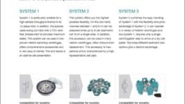
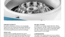
This brochure provides information on the Hettich Cyto System rotors and accessories. (more…) read more...

This product sheet provides a detailed overview of the product features, benefits and models. (more…) read more...
Hettich manufactures centrifuges for any standard laboratory application.